Benefits Of Spine Mri: The Complete Guide
- - Category: Diseases & Conditions
- - 11 Nov, 2024
- - Views: 83
- Save

Magnetic resonance imaging (MRI) is one of the most modern and precise diagnostic technologies actively used in medicine
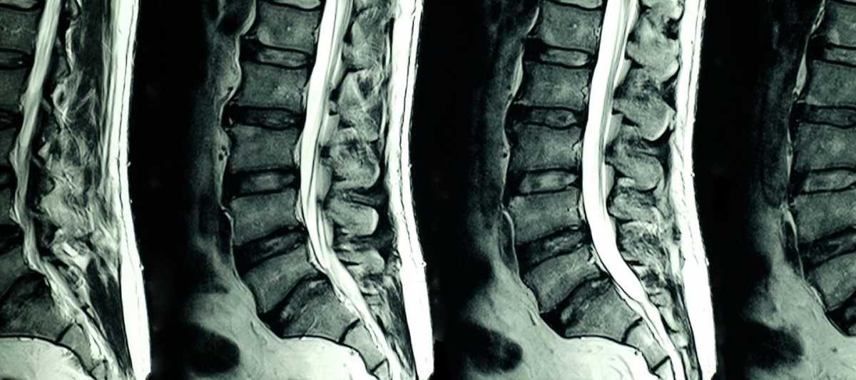
Magnetic resonance imaging (MRI) is one of the most modern and precise diagnostic technologies actively used in medicine. A special role is played by MRI of the spine, which enables a very detailed examination of the spinal cord, intervertebral discs, ligaments, and surrounding structures without invasive procedures. This technique allows doctors to detect various conditions, from degenerative disc disease to tumors, even in the earliest stages of their development.
Basics of MRI and how it works
Unlike X-ray techniques that use ionizing radiation, MRI relies on the effect of a magnetic field and radio frequency pulses on hydrogen atoms in body tissue. MRI produces a strong magnetic field that forces protons in the body to align with that field. When the atoms return to their original condition, they emit signals that are picked up by the devices and converted into detailed images.
The main advantage of MRI is that it can create three-dimensional images of various tissues without using harmful radiation. This is especially important for examining soft tissue that is not always visible on X-rays or computed tomography (CT).
Main benefits of spinal MRI
1. High diagnostic accuracy
One of the main advantages of MRI of the spine is the high accuracy and detail of the images received. This is especially important when examining complex structures such as the spinal cord, intervertebral discs, and nerve roots. MRI can detect even small changes in the tissue and is therefore an essential diagnostic method. Thanks to its high accuracy, MR helps doctors make the right decision regarding treatment approaches, whether it is conservative treatment or surgical intervention.
2. Safety
One of the main advantages of spine MRI is its safety. Unlike X-ray and CT, which use ionizing radiation, MRI doesn’t expose the patient to radiation. This makes it an excellent method for frequent examinations, especially for patients who require long-term follow-up, for example with chronic spine diseases. The procedure can also be recommended for pregnant women (except for the first trimester) and children, which expands the scope of application.
3. Excellent soft tissue visualization
MRI offers extraordinary opportunities for soft tissue imaging. This is important for examining discs, ligaments, nerve roots, and spinal cord. Structures such as muscles and ligaments often play a key role in spinal cord disorders and cannot always be visualized using conventional diagnostic methods such as X-rays. Magnetic resonance, for example, can accurately show the extent of nerve damage caused by a herniated disc, which is important for choosing the right treatment method.
4. Multi-plane images
MRI provides images in multiple projections, making it possible to examine the spine from different angles. This is especially important when assessing the complex structures of the spine and spinal cord, where damage can occur in multiple locations at the same time. For example, a doctor can look at transverse, sagittal, and coronal sections to get a complete picture of the spine.
5. Early diagnosis of diseases
MRI makes it possible to detect diseases in the earliest stages of their development. This is especially important in pathologies such as spinal tumors, inflammatory processes, or infections, where early diagnosis can significantly increase the chances of successful treatment. In addition, MRI helps detect degenerative changes in intervertebral discs and joints before obvious clinical symptoms appear, so that treatment can begin before serious complications occur. If you search for “cervical spine MRI near me”, be sure to choose an experienced and professional team of specialists.
6. Lack of invasiveness
MRI is a non-invasive diagnostic method that doesn’t require surgery or contrast agents (in most cases). This is especially important for patients at high risk of complications from invasive procedures. In rare cases, it may be necessary to inject a contrast medium to make blood vessels or tumors more visible. However, most studies are carried out without this option.
7. Option to take the exam without prior preparation
The patient doesn’t need any special preparation to perform an MRI of the spine. This convenience is especially important in emergencies where a quick diagnosis is required. The patient doesn’t need to fast, take special medication, or undergo complex preparatory procedures, which makes the examination as accessible as possible.

